.svg)
The Complete Shade-Taking Guide for Dental Restorations
Shade selection is the single most common source of remakes in crown and bridge work—not because clinicians lack skill, but because the process is treated as an afterthought. Rushed at the end of a prep appointment, under harsh operatory lighting, on a dehydrated tooth. The restoration itself may be technically flawless, but the mismatch is already built in before the case ever reaches the laboratory.
At Flora Orthodontics, we process hundreds of crown and veneer cases monthly from dental practices across London and the UK. The cases with the highest first-try success rates share one factor: systematic documentation. Dentists who follow a structured shade-taking protocol incorporating proper lighting, value-first selection, material-specific considerations, and standardised photography experience significantly fewer remakes.
This guide presents a practical, seven-step protocol developed specifically for dentist-to-laboratory communication. Whether you are working with traditional analog shade guides or digital shade-matching devices, these principles will improve your restoration outcomes.
The Three Dimensions of Tooth Colour
Tooth colour is not a single variable. It operates across three independent axes, and misreading any one of them produces a mismatched restoration.
Understanding Hue, Value, and Chroma
Hue is the base colour family the quality that distinguishes a reddish-brown tooth from a greyish one. On the VITA Classical guide, hue is encoded by the letter prefix: A (reddish-brown), B (reddish-yellow), C (grey), D (reddish-grey). Most natural teeth fall within the A and B ranges.
Value is brightness how light or dark the tooth appears regardless of its hue. Value is the most critical dimension in anterior aesthetics. A restoration that is correct in hue but too low in value will read as obviously artificial against its neighbours. When in doubt, go one step higher in value. A slightly brighter result is correctable; a darker one is not.
Chroma describes the saturation or intensity of the hue. The numerical suffix on a shade tab indicates chroma level: 1 is low saturation, 4 is high saturation. Chroma increases from the incisal to the cervical zone, which is why a single shade tab never fully describes a natural tooth.
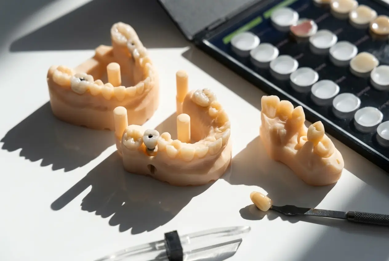
Shade Guide Systems Compared
Not all shade guides work the same way. Understanding the differences between the two dominant systems is essential for consistent lab communication.
Key Point: Mismatches arise frequently when the dentist uses a VITA Classical tab label but the lab technician references a 3D-Master equivalent. Always confirm which system your laboratory works from before prescribing.
The 7-Step Shade-Taking Protocol
Step 1: Timing Select Shade Early
Shade selection must occur at the start of the appointment, before any preparation or retraction that will dehydrate the enamel. A dehydrated tooth appears 1–2 value steps lighter than its true resting state. If shade is selected after prep, the restoration will be fabricated too bright and will visibly darken within 24 hours of cementation as the tooth rehydrates.
Step 2: Control the Environment
Remove bright lipstick. Cover vividly coloured patient clothing with a neutral grey bib. Ensure your gloves are neutral-coloured rather than bright latex. Position the patient upright at eye level, approximately 25–35 cm away.
Turn off your operatory light—its intensity creates glare and alters perceived colour. Ideally, conduct shade selection near a north-facing window with natural daylight (colour temperature 5500–6500K), which provides the most accurate colour rendering.
Step 3: Determine Value First
Using your shade guide, identify the lightness level first. Squint your eyes to reduce colour perception and focus on brightness. The VITA 3D-Master system simplifies this with its value-based organisation (levels 0–5).
Record the value level clearly. If between values, always select the lighter option—extrinsic characterisation can darken a restoration, but lightening a too-dark crown requires remaking.
Step 4: Select Hue and Chroma
Once value is established, select the appropriate hue (A, B, C, or D) and chroma level. Hold the shade tab parallel to the tooth surface at the same plane—positioning it behind makes it appear darker; in front makes it appear lighter.
Limit each comparison to 5–7 seconds maximum. Prolonged staring fatigues your cone cells and reduces colour discrimination. Between comparisons, rest your eyes by focusing on a neutral grey or blue surface to resensitise your vision.
Step 5: Map Shade by Tooth Thirds
Natural teeth rarely present a single uniform shade. Document each third separately:
- Cervical third: More chromatic, warmer tone
- Middle third: Base shade selection
- Incisal third: Translucency, halo effects, opalescence
Include notes on surface texture, lustre, and unique characteristics such as white spots, crack lines, or staining patterns.
Step 6: Photograph with Reference Tab
Take at least three photographs:
- Full smile view — restoration in context with surrounding dentition
- Close-up with shade tab — tab positioned adjacent to target tooth, parallel to surface
- Close-up without tab — captures surface characteristics and translucency
Use a macro lens with ring flash for consistent, shadow-free lighting. Include a shade tab in every photograph—it provides the technician with colour calibration reference.
Step 7: Verify Under Multiple Light Sources
Verify your shade selection under different lighting conditions. If the match holds under both natural daylight and fluorescent operatory light, you have minimised metamerism risk.
Material-Specific Shade Considerations
Shade taking is not material-agnostic. The prescription changes depending on what the technician will fabricate. Understanding how different materials transmit, reflect, or block light is essential for accurate outcomes.
Critical Note: For IPS e.max cases, failing to communicate the stump shade is the leading cause of unexpected grey shadowing visible through the restoration. Always include a stump shade tab photograph for translucent ceramic cases.
Digital Shade Matching: When to Invest
Spectrophotometers and colorimeters remove subjectivity by measuring reflected light across wavelengths and mapping results to specific shade tab references. These devices are consistent, repeatable, and unaffected by observer fatigue or ambient lighting.
Consider adding digital shade matching if:
- You frequently handle high-aesthetic anterior cases
- Your practice struggles with consistent shade-related remakes
- You work with multiple laboratories and need standardised communication
- Patients present challenging shade characteristics (high translucency, complex characterisation)
Popular devices include VITA Easyshade, ShadeScan, and SpectroShade. While initial investment is significant, practices handling substantial aesthetic work often recover costs through reduced remakes.
Remember: Digital devices supplement, not replace, clinical judgment. The most reliable approach combines digital readings with traditional shade guide verification and comprehensive photography.
Common Mistakes (And How to Avoid Them)
Mistake 1: Relying solely on the shade tab number
The shade tab indicates a range, not an exact match. Always supplement with value assessment, photography, and descriptive notes.
Mistake 2: Shade selection after preparation
Dehydrated teeth and irritated soft tissues distort colour perception. Select shade before you begin tooth preparation.
Mistake 3: Ignoring the cervical third
The cervical third often requires different characterisation than the middle third. Failing to document this leads to mismatched emergence profiles.
Mistake 4: Inadequate photographs
Blurry, poorly lit, or poorly composed photographs force technicians to guess. Take the extra minute to capture sharp, properly exposed images with shade tabs.
Mistake 5: Not accounting for metamerism
Verify shade matches under at least two different light sources to minimise colour mismatches in different environments.
Mistake 6: Omitting material specification
Different ceramics require different shade communication. e.max needs stump shade; zirconia needs ingot specification.
What Your Dental Lab Needs From You
The restoration is only as accurate as the information sent with the case. A complete shade prescription should include:
- The shade guide system used (VITA Classical or 3D-Master)
- Body shade, cervical shade, and incisal shade as separate values
- Stump shade for all translucent ceramic cases (e.max, layered zirconia)
- Material-specific notes (characterisation, high translucency, bleach shade target)
- A calibrated clinical photograph with a shade tab in frame
- Patient age (enamel translucency and surface texture change significantly with age)
At Flora Orthodontics, detailed shade documentation is the baseline expectation for every case. Remakes cost everyone time and money. The cases that arrive with complete, structured shade prescriptions are the ones that pass at first fit.
Download Free Shade Prescription Template
Frequently Asked Questions
When should shade selection occur during a dental appointment?
Always at the beginning, before preparation or dehydration. Dehydrated enamel appears 1–2 value steps lighter, causing restorations to darken visibly after cementation.
Why do crowns sometimes look different despite matching the shade tab?
Material opacity, underlying stump shade, lighting conditions, and incomplete lab communication all affect final appearance. A shade tab is a reference, not a guarantee.
Is a stump shade always necessary for crown cases?
Essential for translucent ceramics like e.max where underlying colour shows through. Less critical for opaque zirconia or PFM restorations that mask the preparation.
How long should each shade comparison take?
Limit to 5–7 seconds maximum. Prolonged staring causes eye fatigue and reduces colour discrimination. Rest eyes on neutral grey between comparisons.
Are digital shade-matching devices better than visual guides?
They reduce subjectivity and provide repeatable data, but supplement rather than replace clinical judgment. Best results combine digital readings with visual verification.
What causes metamerism in dental shade matching?
Metamerism occurs when two objects match under one light source but differ under another. Always verify shades under natural daylight and operatory lighting.
Why is value more important than hue in shade selection?
Value errors are immediately visible; moderate hue inaccuracies often go unnoticed. A bright wrong hue looks more natural than a correct hue that is too dark.
Conclusion
Accurate shade taking is a clinical protocol, not a clinical talent. It follows rules: take the shade early, in the right light, without eye fatigue, with material-specific precision, and with complete documentation. The output of that protocol—a structured, complete prescription—is what allows the laboratory to produce work that integrates seamlessly into the patient's dentition.
The gap between a good restoration and a failed one is almost never the ceramics. It is the information that accompanied the case.
For dental practices in London seeking a laboratory partner that understands the importance of clear communication, Flora Orthodontics offers same-day case confirmation, digital workflow support, and technician consultation for complex aesthetic cases.
Submit Your Next Case to Flora Orthodontics | Download Shade Mapping Template
Flora Orthodontics is a full-service dental laboratory serving dental practices across London and the UK. We specialise in crown and bridge restorations, aesthetic veneers, and complex aesthetic rehabilitations with industry-leading turnaround times.
Frequently Asked Questions
Our team answer to your questions
Always at the beginning, before preparation or dehydration. Dehydrated enamel appears 1-2 value steps lighter, causing restorations to darken visibly after cementation.
Material opacity, underlying stump shade, lighting conditions, and incomplete lab communication all affect final appearance. A shade tab is a reference, not a guarantee.
Essential for translucent ceramics like e.max where underlying colour shows through. Less critical for opaque zirconia or PFM restorations that mask the preparation.
Limit to 5-7 seconds maximum. Prolonged staring causes eye fatigue and reduces colour discrimination. Rest eyes on neutral grey between comparisons.
They reduce subjectivity and provide repeatable data, but supplement rather than replace clinical judgment. Best results combine digital readings with visual verification.
Metamerism occurs when two objects match under one light source but differ under another. Always verify shades under natural daylight and operatory lighting.
Value errors are immediately visible; moderate hue inaccuracies often go unnoticed. A bright wrong hue looks more natural than a correct hue that is too dark.