.svg)


.svg)
Crown and Bridge Dental Laboratory for UK Dentists
Most dentists do not change crown and bridge laboratory because the fabrication is poor. They change because of everything around the fabrication the crown that needs adjusting at every fit, the bite that was never quite right, the case that comes back a week later than promised, the shade query that goes unanswered. The technical work might be competent. The relationship is not.
Flora Orthodontics is a crown and bridge laboratory for dental practices across London and the UK. We work in zirconia, e.max, PFM and gold, across both digital and conventional workflows, and we are built around a single operating principle: the restoration that fits at first appointment is decided long before it is milled or pressed. It is decided by how the material was selected, whether the preparation matched it, and what information reached the laboratory with the case.
This page sets out how we work what we make, the materials we use and when, how we handle digital and conventional cases, our turnaround and communication, and the records that let us return work you can seat without a tray full of adjustment burs.
What Our Crown and Bridge Service Covers
Flora provides the full fixed-prosthetic range a restorative practice needs from a single laboratory:
- Single crowns — monolithic and layered zirconia, e.max, PFM, and full-cast gold
- Conventional bridges — including Maryland (resin-bonded) and cantilever designs
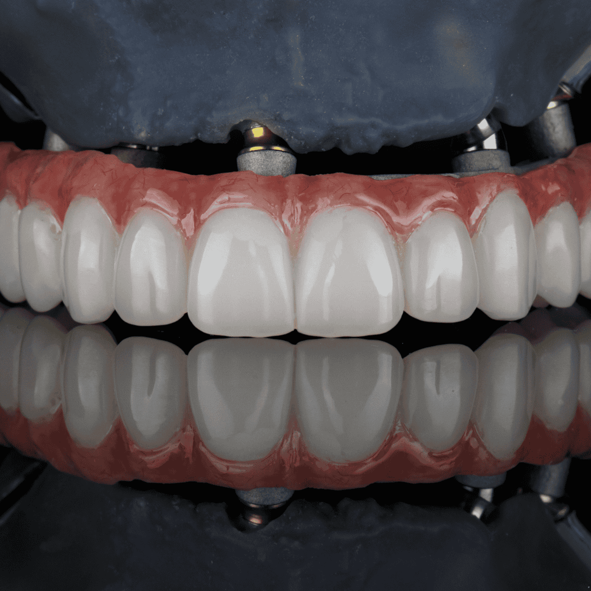
- Implant crowns and bridges — screw-retained and cement-retained, on the major implant systems
- Inlays and onlays — for cases beyond a direct restoration but short of a full crown
- Post and cores — cast and prefabricated, to restore endodontically treated teeth before crown work
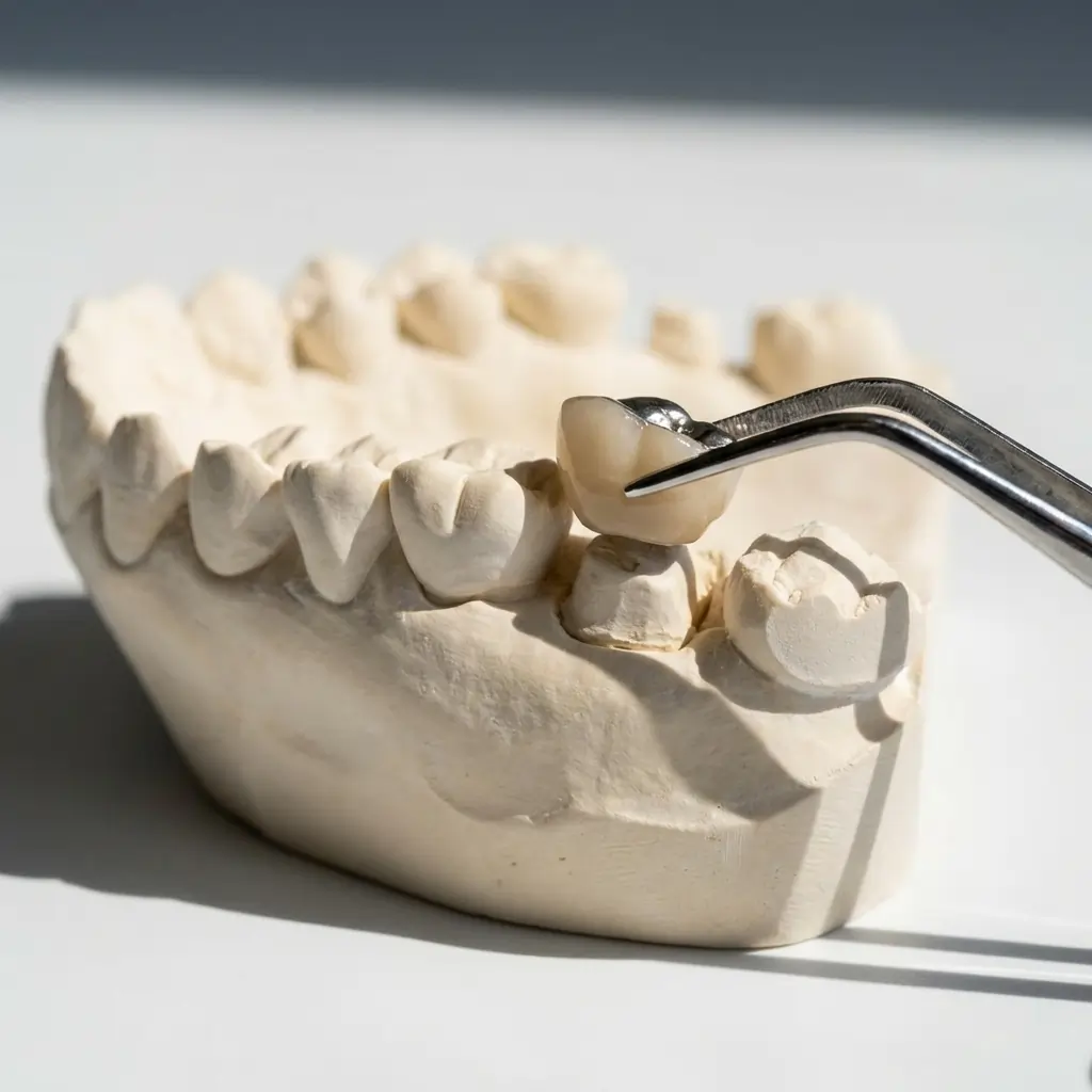
Every case is fabricated to prescription from your impression or intraoral scan, by GDC-registered dental technicians, and returned ready to fit.
The Real Reason Crown and Bridge Cases Go Wrong
Most crown and bridge problems are locked in before fabrication begins. The three most common causes are not technical failures — they are information gaps.
Insufficient preparation space. Every crown material has a minimum reduction requirement. Zirconia, e.max, PFM and gold each behave differently under load and require different thicknesses to function. Prescribing a material without verifying that the preparation can accommodate it produces a crown that is either too thin to be reliable or too bulky to fit passively.
Unreliable bite records. A bite taken after a long anaesthetic, with the patient partially open, or across too many teeth produces an inaccurate mounting. The restoration seats on the model but not on the patient. Occlusal adjustment follows, and the margin of that adjustment determines whether the case works or has to be redone.
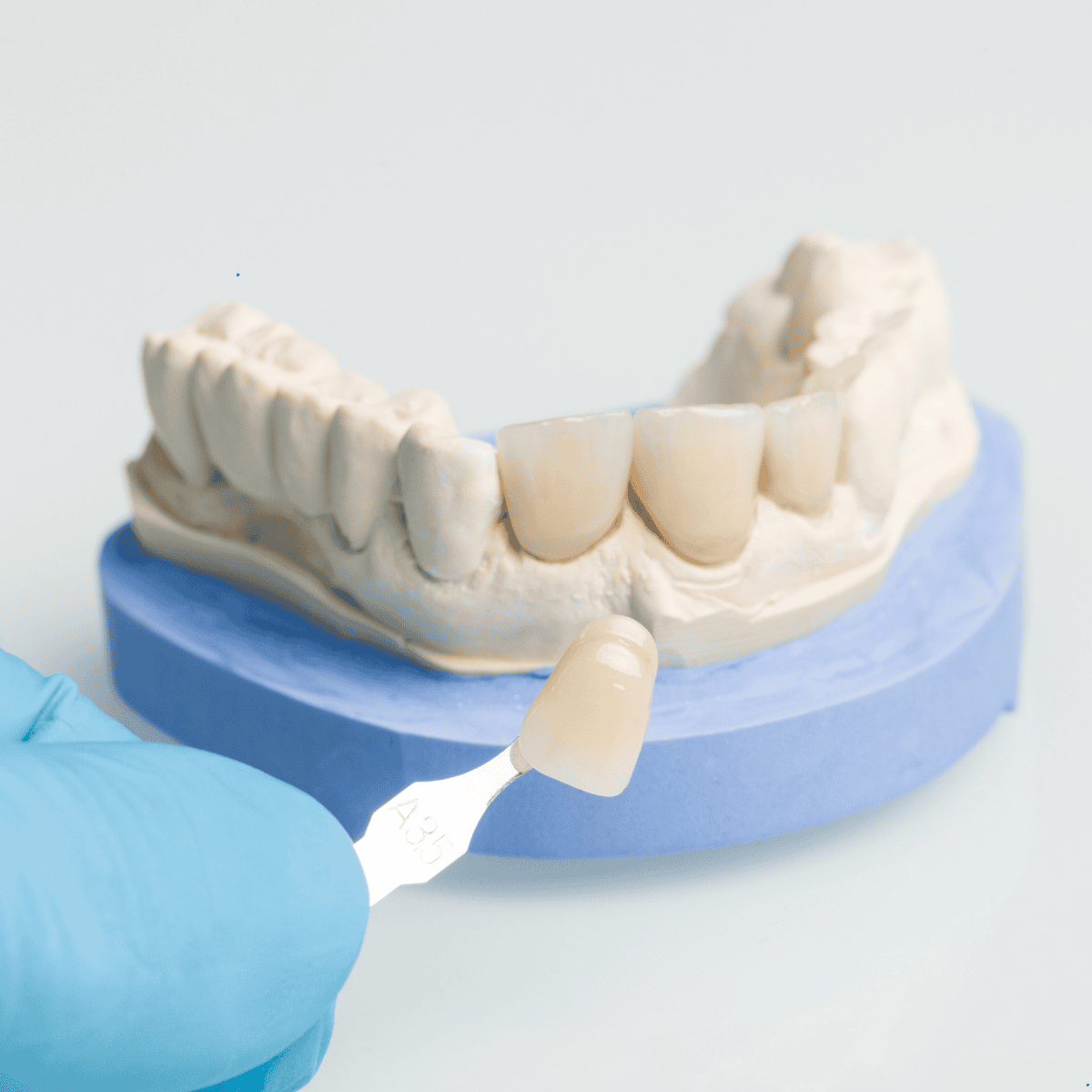
Inadequate shade communication for visible cases. A single shade code, without photography, stump shade or notes on the adjacent teeth, is not shade communication. It is a starting point the technician must interpret. In highly visible anterior work, that interpretation gap is where aesthetically disappointing crowns begin.
A laboratory cannot fix these after the fact. What it can do is flag them before fabrication — which is the difference between a lab that processes prescriptions and a lab that protects your chair time.
Choosing the Right Crown Material for the Case
Material selection should follow the clinical objective, not the reverse. In practice, that means asking four questions before the preparation begins:
- What is the primary demand aesthetics, strength, or both?
- How much space does the preparation realistically allow?
- Where is the restoration in the arch, and will the patient show it on smile?
- Does this patient have parafunctional habits that affect material durability?
The minimum reduction figures above are not arbitrary. They are the thresholds below which the material cannot reliably perform its intended function. A zirconia crown fabricated under 1.0 mm will be over-contoured to achieve the required strength; an e.max crown prepared to 1.0 mm occlusal reduction will fracture under posterior load.
For the detail behind each material, see our deep-dive guides on e.max crowns, zirconia crowns and PFM and gold restorations.
Zirconia, e.max, PFM and Gold When Each Is the Right Choice
Zirconia Crowns
Zirconia has become the dominant posterior crown material in most active practices, and for good reason. Monolithic zirconia is strong, efficient to fabricate and highly predictable when the preparation space is adequate. It does not depend on shade photography for posterior functional cases, and it tolerates the parafunctional forces that ceramic restorations handle less reliably.
We distinguish between two clinical applications. Monolithic zirconia is the right choice when the objective is posterior durability and occlusal stability. Layered zirconia becomes relevant when the unit is visible and you need improved optical integration — but layered cases require the same shade documentation investment as an e.max case. Prescribing layered zirconia with a single shade code and no photography produces a result that is better than monolithic for aesthetics but still limited by the information provided.
e.max Crowns
e.max remains one of the strongest choices available for anterior aesthetic work. Its translucency and optical depth, combined with accurate shade documentation, can produce restorations that are genuinely difficult to distinguish from the adjacent natural teeth.
That outcome requires investment at the chair. The e.max cases that deliver outstanding anterior aesthetics consistently share the same inputs: shade taken before preparation on a hydrated tooth, clinical photography with a shade tab, a stump shade where the preparation shows through, and characterisation notes for cervical chroma and incisal translucency. Without those inputs, the laboratory is making artistic decisions on incomplete information. For posterior e.max, the material is appropriate when adequate space exists and the case is not high-load; under 1.5 mm of posterior clearance, monolithic zirconia is the more predictable choice.
PFM Crowns
PFM is no longer the universal posterior crown, but it remains clinically appropriate where the unit is not aesthetically demanding, the case is cost-sensitive, or a metal-supported restoration is preferred for a specific patient. The most common problem with PFM prescriptions is over-reduction at the metal collar, which creates a thin margin that chips early — the preparation geometry needs to account for the shoulder or chamfer at the porcelain margin and the space for the metal substructure.
Gold and Full-Metal Crowns
Gold crowns are underused relative to their clinical merits. Full-metal restorations offer the most conservative preparation of any crown type, excellent precision of fit, and a long clinical track record that modern ceramics are still building toward. Where the tooth is not visible and the primary objective is functional longevity, no modern ceramic reliably outperforms it. Flora supports full-metal crown and bridge work as part of a complete laboratory offering.
Bridge Cases Need More Than a Material Label
A prescription that says "3-unit zirconia bridge" has communicated the span and the material. It has not communicated the design — and bridge design decisions significantly affect how the case functions, how the patient maintains it, and whether we can fabricate it correctly the first time.
Before a bridge case is submitted, the following should be defined:
- Pontic form. Modified ridge lap, ovate and sanitary pontics are not interchangeable. Ovate pontics require a prepared tissue site; modified ridge lap is the most common for posterior function; sanitary reduces maintenance in non-visible positions but is rarely ideal aesthetically.
- Connector dimensions. Connectors carry the entire load across the span. Under-designed connectors fracture — which is why minimum cross-sections matter: typically 9 mm² for posterior zirconia spans and 16 mm² for e.max. These are fracture thresholds, not preferences.
- Span length. Longer spans change stress distribution and affect material selection. Multi-unit spans that test e.max's connector requirements may be better suited to zirconia or PFM.
- Tissue relationship and hygiene access. If the pontic space has significant ridge resorption, the aesthetic and hygienic management of the area should be stated, not assumed.
The bridge cases that produce difficult conversations at fit are the ones where the design was inferred from the models because the prescription did not specify it.
Want to learn more about bridges and partial check our article here
Digital and Conventional Workflows Both Done Properly
Flora works in both digital and conventional workflows, and a case is not better or worse for the route it takes. What matters is that the records are complete for whichever route you use.
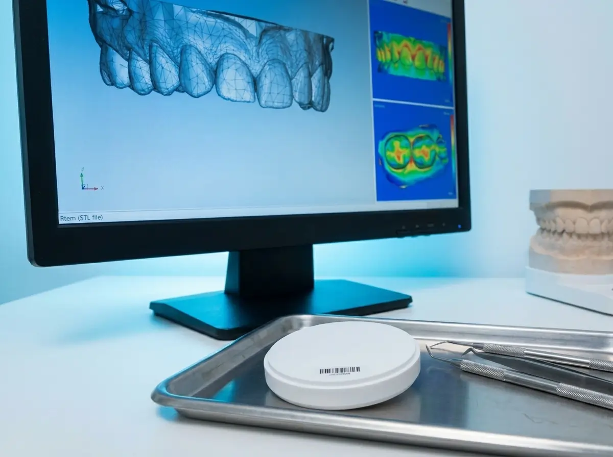
Digital cases. We accept open STL files from the major intraoral scanners — iTero, 3Shape TRIOS, Medit, Dexis and Primescan among them. For a digital crown we need the preparation scan, the opposing arch scan and a buccal bite scan; the preparation scan must capture the complete margin circumferentially, because a partial or noisy scan of the margin is the single most common cause of digital fit issues. Designs are milled or pressed and finished — stained, glazed and fired — to the prescribed shade and strength.
Conventional cases. We use definitive PVS or polyether impressions for all conventional crown and bridge work. Alginate is appropriate for study models and opposing arches in routine posterior work, but not for the working impression — it distorts during casting and produces margins we cannot work from confidently.
Working With Flora Turnaround, Coverage and Communication
A laboratory relationship is judged on three things beyond the restoration itself: how quickly work comes back, how reliably it arrives, and whether you can speak to the person making it.
- Turnaround. Crown and bridge work is returned on a predictable schedule, with same-day and next-day options for time-critical cases in and around London and nationwide.
- UK-wide coverage. delivery across London and the UK, with daily collection in central London.
- GDC-registered technicians. Every restoration is made by registered dental technicians working to documented quality standards.
- Technician access on complex cases. For multi-unit aesthetic work, implant cases or difficult shade matches, you can speak directly to the technician handling the case before it is fabricated — not after it has been remade.
The point of all of this is fewer remakes and fewer adjustment appointments. A laboratory that returns work you can seat predictably is worth more to a busy practice than a marginally cheaper unit price that costs you chair time at every fit.
The Case Information That Most Reduces Adjustments
Most avoidable chairside adjustments trace back to one of five information gaps at submission:
- Reduction not verified before prescription. The material was chosen for the right reasons, but the preparation space was not measured — and the crown comes back over-contoured because there was nowhere to put the required material volume.
- Bite record taken after extended preparation. Once teeth are prepared and anaesthetised, the patient loses proprioceptive reference for intercuspal position. Take the bite with minimal material, over the prepared and adjacent teeth, with the patient actively closing to maximum intercuspation.
- Bridge prescribed without pontic instructions. Underdefined pontic design is the most consistent source of bridge adjustment conversations. If the form is not specified, we make a reasonable default decision — which may not match your intent.
- Anterior case submitted without photography. In posterior work, shade code alone is usually sufficient. In anterior work, even basic photographs in good light reduce mismatches significantly.
- Stump shade not documented for e.max cases. When the preparation is discoloured or dark, the absence of a stump shade note often produces an opaqued restoration that reads flat and artificial.
A complete submission includes restoration type and number of units; material and specific type; arch, tooth numbers and abutment details for bridges; the working impression or STL files; shade note with photography for visible units; stump shade where relevant; and pontic form, connector intent and tissue notes for bridge cases.
Shade communication is the highest-leverage habit in the whole process — our shade-taking guide for dental restorations sets out the seven-step protocol we recommend.
How to Send Your First Case to Flora
Starting with Flora does not require changing how you work:
- Tell us your workflow. Whether you scan or impression, and which system you use — we accept open STL from all major scanners and definitive conventional impressions.
- Send a first case. A single posterior crown or a straightforward anterior unit is the usual way practices assess fit, contacts and aesthetics.
- Build a profile. We record your preparation preferences, preferred materials and margin style so repeat work is consistent to your practice.
Want to learn more ?
e.max Crowns
When to prescribe lithium disilicate, the preparation it needs, ingot selection and the cementation protocol. A clinical guide for dentists.
Zirconia Crowns
The 3Y/4Y/5Y strength-and-translucency trade-off, the three restoration types, prep guidelines and why zirconia cements differently from e.max.
Dental Bridges
Which material, for which case. Strength, aesthetics, preparation and cementation.
Frequently Asked Questions
Our team answer to your questions
Monolithic zirconia typically requires 1.0–1.5 mm of occlusal reduction and 0.5–0.8 mm of axial reduction. Layered zirconia cases need slightly more space at least 1.5 mm occlusal to accommodate the ceramic layer without over-contouring the substructure.
e.max is typically the better choice for anterior single units and other visible cases where translucency and optical depth are priorities. It requires a minimum of 1.5–2.0 mm occlusal reduction and returns the best aesthetic results when paired with thorough shade documentation and clinical photography. For high-load posterior cases or limited preparation space, monolithic zirconia is usually more appropriate.
Yes. PFM and full-metal crowns remain clinically appropriate in selected cases and form part of Flora's complete crown and bridge offering. Full-metal restorations in particular offer the most conservative preparation of any crown type and excellent long-term durability in posterior non-aesthetic positions.
We need the full preparation scan, opposing arch scan, and a buccal bite scan at minimum. For anterior or aesthetic cases, clinical photographs are strongly recommended. The preparation scan must capture the complete margin circumferentially — partial or noisy scans of the margin area are the most common cause of digital crown fit issues.
The three most common options are modified ridge lap (the most common posterior choice), ovate (requires a prepared tissue site, best aesthetics), and sanitary (easiest hygiene access but least aesthetic). If no instruction is given, the laboratory will select a default form that may not match the clinical intent — particularly for visible anterior bridges where the aesthetic objective should be stated explicitly.
Connectors carry the load across the entire span. Under-designed connectors are the most common cause of bridge fracture. For zirconia bridges, minimum connector cross-section is typically 9 mm² for posterior spans; for e.max, 16 mm². If the preparation space or case design does not allow adequate connector dimensions, the material choice should be revisited before fabrication begins.
A shade code tells the laboratory which tab to reference. A photograph shows them the actual optical environment the restoration needs to integrate with — the value, translucency, surface texture, and cervical chroma of the adjacent teeth. For anterior aesthetic cases, photography is the most effective single action a practice can take to reduce the probability of a shade-related remake.